Z02: Visualization of Biopolymer Function and Research Data Management
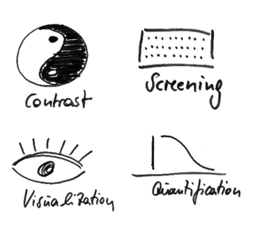
Whereas in vitro studies (here referring to studies using purified components) can provide molecular insights, in vivo model systems (here also referring to studies within cells) are the minimal means by which to study biomolecular function within physiological contexts. Non-invasive microscopy methods have proven valuable for in vivo work and, if the complexes are large, even in vitro. In addition, microscopy and spectroscopy methods are frequently related, or can be performed using the same equipment, such that even single molecules can be studied using fluorescence microscopes. For the proposed CRC, we will establish an innovative service platform for quantitative microscopy. The platform will be composed of four pillars comprising state-of-the-art microscopy techniques for i) static and ii) dynamic investigation of polymer dynamics and assembly processes, iii) innovative projects in which we will adapt label-free imaging techniques, and iv) knowledge transfer. The output, from the service project Z02, that is, images, videos and quantitative parameters will be collated in a common database, accessible by all partners. This project will also coordinate and undertake all efforts related to research data management (RDM) across the whole CRC.
Parameters and microscopy techniques provided by LMCF-BIO, MHCF-IMB and our collaboration partners at the MPI-P within the Z02 service project.
Bastian B. Hülsmann
Faculty of Biology Light Microscopy Core Facility, JGU